You must be signed in to read the rest of this article.
Registration on CDEWorld is free. You may also login to CDEWorld with your DentalAegis.com account.
When restoring the worn dentition, it is absolutely critical to establish proper function of the masticatory system. In many cases, when patients present with worn or fractured teeth requiring restorations, clinicians should do a thorough examination to determine a diagnosis and cause of the current dental condition with which the patient presented. Dental occlusion has been such a widely discussed and debated topic, perhaps creating confusion that may limit the implementation of ideal occlusal principles into many everyday practices.1,2 While beautiful restorations can be fabricated with the excellent materials available today, the longevity of these restorations and the stability of the chewing system may be significantly impacted if these restorations are not placed in an ideal occlusal environment.3 In some cases, an ideal situation is often unattainable due to tooth position, structural integrity of the teeth, skeletal issues, or the desires of the patient. In these cases, as will be discussed in the case presentation, compromises are made but it is still important to create an occlusal environment that is as stable as the situation dictates. These compromises can be made in the restorative materials used or in the use of an occlusal splint.
Comprehensive treatment planning should always begin with a thorough examination and obtaining a complete patient history. This examination should include a review of the patient’s medical and dental history, along with a conversation with the patient discussing the expectations of treatment. Clinically, a review and diagnosis of the necessary radiographs, along with a detailed periodontal assessment and oral cancer screening, should be completed. The functional portion of the examination should include palpation of the muscles of mastication, a mandibular range of motion test, and a thorough evaluation of the temporomandibular joints, including load testing and Doppler auscultation of the joints. An evaluation of the airway and a sleep apnea risk assessment is also performed. Finally, the dentition should be examined for signs of wear, tooth fracture, mobility, migration, and decay.
Following the complete examination, a review of findings appointment is scheduled, allowing the dentist and patient to discuss the problems, the implications of those problems, and to go over treatment possibilities.4 If the patient decides to ideally address their issues in a comprehensive manner, proper records are then taken, which include diagnostic photographs and models, a centric relation bite record, and a facebow transfer. This information will then be used to determine a precise treatment plan that will address the patient’s concerns while providing ideal esthetic and functional results in a predictable manner.
Proper Function of the Masticatory System
Joint Position and the Muscles of Mastication
Function of the masticatory system begins with the temporomandibular joints.5 The joints should be healthy, free of pathology and degeneration, and should ideally be positioned in centric relation. Dawson defines centric relation as the relationship of the mandible to the maxilla when the properly aligned condyle-disc assemblies are in the most superior position against the eminentiae irrespective of vertical dimension or tooth position.6
It is extremely important for the reader to understand that centric relation is totally independent of tooth position. It is solely related to the position of the joints. Also, centric relation is not a position that a patient is “forced” or “put” into. It is a natural anatomic position that is achieved by normal coordinated contraction of the elevator muscles. This precise position can be recorded and reproduced with pinpoint accuracy using Dawson’s bimanual manipulation technique.7 As the elevator muscles work to close the mandible, the condyles are naturally positioned within the glenoid fossa. Without influence from occluding teeth, the contraction of the masticatory muscles seat the condyles into centric relation.8 Once the condyles are in centric relation, the lateral pterygoid muscles are allowed to release and the elevator and depressor muscles of mastication can function in harmony with one another.9,10 This harmonious activity significantly reduces the development of temporomandibular joint disorders, muscle pain, and many other associated issues. When the joints are not allowed to achieve centric relation, the elevator and depressor muscles begin working against one another, resulting in an increased possibility of muscle pain, joint pain, and other signs and symptoms of occlusal instability.11
As stated before, centric relation is not a forced position. Many dentists feel that the bimanual manipulation technique is a way to force the patient’s mandible into centric relation. This is not true. Bimanual manipulation should only be used to verify that the patient is in centric relation by assessing the level of tension or tenderness within the joint upon loading. It is the author’s opinion that the term “bimanual manipulation” would be better phrased as “bimanual verification” in order to alleviate much of the confusion regarding this extremely important and necessary technique.
Tooth Position and Proper Occlusal Contacts
Once again, centric relation is based solely on the position of the condyles within the glenoid fossae. Once the condyles are properly seated, the position of the teeth and occlusal contacts should be idealized so as to not interfere with centric relation. This means that as the mandible hinges into a closed position, all of the teeth should contact simultaneously in order to eliminate a premature tooth contact resulting in a slide of the mandible. Any sort of hit-and-slide from centric relation into maximum intercuspation will cause the condyles to translate down and forward out of the fossae. Once the condyles are positioned down and forward on the slippery slope of the eminentiae, the inferior belly of the lateral pterygoid muscle must contract to hold the condyles in this down and forward position, while the superior belly of the lateral pterygoid must also contract to keep the disc properly positioned between the condyle and eminence. 12-14 Periods of prolonged contraction of the lateral pterygoid result in fatigue or spasm of the muscle, which can be experienced as pain and discomfort to the patient.15,16 These symptoms can be exacerbated if the patient has a clenching or bruxing habit because the temporalis, medial pterygoid, and masseter elevator muscles will be highly active and will be in direct contrast to the already contracted lateral pterygoid muscles.17 This dysfunction and constant opposition between the elevator muscles and condyle positioning muscles will further increase the fatigue and strain on all of the muscles of mastication.
Also, constant tension within the superior belly of the lateral pterygoid muscle (the portion of the lateral pterygoid with attachments to the articular disc) will result in continuous stretching of the ligaments that attach the disc to the posterior surface of the condyle. This constant stretching can eventually create an unstable condyle-disc assembly, resulting in a disc that can click or pop off of and onto the lateral pole of the condyle during function (Figure 1).18 Further damage can be created if the medial aspect of the disc slides forward and off of the condylar head. This severe situation can result in the head of the condyle pressing against the highly innervated and vascular retrodiscal tissue. This can be experienced as extreme pain in the joint whenever the patient attempts to close their mandible or bite into food. If left untreated, this could eventually lead to bone-on-bone contact between the head of the condyle and the eminence and potential degeneration of the condylar head.19
Once the teeth come into contact, an ideal cusp-tip-to-fossa relationship should be created between the upper and lower posterior teeth. Contacts on inclines of the teeth must be avoided. This eliminates the possibility of a mandibular shift or noxious lateral forces on teeth that could result in tooth mobility, fracture, wear, pain, sensitivity, or abfractions of the teeth.20 Light contact between the lower incisal edges and upper cingulum will also provide necessary anterior stops. These contacts must be equal throughout the mouth, ensuring no occlusal overload on any particular tooth.
Canine and Anterior Guidance
As the patient begins to function, canine guidance is created to eliminate any working side and most importantly, balancing side interferences during lateral excursive movements (Figure 2). Presence of interferences will further increase the possibility of hyperactive muscle activity and possible destruction of the teeth and joints.21 Anterior guidance is created to provide immediate separation of the posterior teeth during protrusive movements. When the posterior teeth are separated during functional movements, the amount of force delivered by the elevator muscles is significantly reduced. This natural programming allows for protection of the teeth along with relaxation and harmonious coordination of the muscles of mastication.22,23
The anterior guidance created must be in harmony with the envelope of function. This means that the lingual surface of the maxillary incisors must be steep enough to provide necessary guidance for posterior tooth separation, but shallow enough to provide a comfortable guidance path, and concave enough to allow the mandibular incisal edges to freely move during mastication and speaking. Any interference between these surfaces will result in a restricted envelope of function, which may contribute to restoration failure, wearing of teeth, and patient discomfort.24
Maxillary Incisal Edge Position
Another important aspect of masticatory function is the position of the maxillary incisal edge. This edge must be strategically placed in the proper horizontal and vertical position in space. The inner vermillion border of the lower lip determines the horizontal position. Ideally, the incisal edge should contact this wet-dry line on the lower lip to allow for proper enunciation of “F” and “V” sounds during speech. The vertical position of the incisal edge will determine the length of the tooth and esthetic characteristics of the patient’s smile. A happy medium must be found where the maxillary incisors are long enough to provide proper guidance and fit within the envelope of function, but also provide ideal phonetics and esthetics.25,26
As a former mechanical engineer, the author greatly appreciates the many nuances and intricacies of the masticatory system. Like any other finely tuned system or well-oiled machine, a properly designed and ideally functioning masticatory system will result in long-term dental stability and comfort for the patient.
Case Presentation
Patient History and Examination
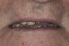
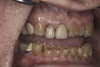
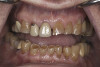
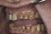
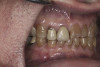
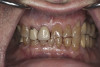
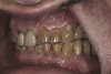
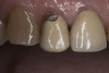
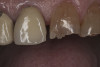
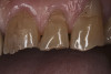
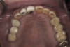
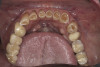
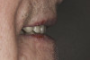
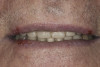
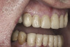
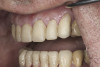
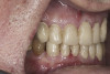
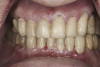
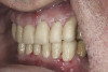
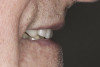
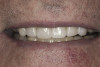
A 77-year-old man presented with severe breakdown of his dentition. A thorough review of his medical history resulted in no contraindications to dental treatment. A discussion of his dental history revealed that the patient was happy with the treatment he had received in the past, but he was interested in “evening out his smile” and ensuring his teeth do not continue to break and chip.
A complete examination was performed. The patient showed no signs of joint dysfunction and there was no tenderness to palpation of any of his muscles of mastication. His mandibular range of motion was within normal limits and his function was completely comfortable. Loading of the joints in centric relation was possible with no signs of tension or tenderness. The first point of tooth contact in centric relation was noted on tooth No. 3 with a 1 mm slide forward from centric relation into maximum intercuspation. Doppler auscultation of the joints showed no signs of clicking/popping or crepitus. Examination of the teeth showed significant wear, abfractions, and erosion into the dentin of the anterior teeth. The patient also had a history of fractured porcelain on his posterior restorations and crowns coming loose. An evaluation of his occlusion revealed that the patient lacked stable centric stops on all teeth. Anterior and canine guidance was non-existent, which led to no separation of the posterior teeth during protrusive and lateral movements. His maxillary and mandibular occlusal planes were also not ideal. The patient reported no airway issues while sleeping and he had a Class 1 Mallampati score. The patient was advised to consult with his physician to evaluate for an acid reflux issue.
Record-Taking and Treatment Planning
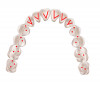
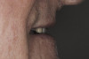
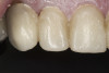
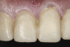
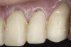
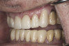
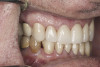
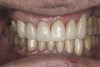
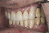
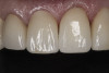
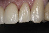
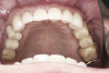
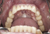
A complete set of records was taken, including diagnostic photographs (Figure 3 through Figure 16), diagnostic impressions, a centric relation bite record, and a facebow transfer. The models were mounted onto an articulator and, along with the photographs, a preliminary workup of the case was completed. An ideal treatment plan consisting of a fullmouth rehabilitation with full-coverage restorations to idealize the planes of occlusion and to properly restore the anterior teeth was presented to the patient. The patient was not interested in extensive work on his posterior teeth because of his age and the fact that he was only interested in “something that will last another 10 years because that’s about how long I’ll be around.” He was only concerned with addressing the anterior teeth and would not accept a treatment plan replacing the restorations on his posterior teeth. Because the author wanted to help the patient with his esthetic concern while still addressing the functional issues, an alternative compromise treatment plan was agreed upon, which involved definitive full coverage restorations in the anterior and composite bonding in the posterior. The patient was fully aware that this treatment plan was a compromise and that the posterior composites will most likely require repair over time. By stabilizing the anterior contacts, anterior guidance, and canine guidance, the author felt comfortable placing bonded composite restorations on the posterior teeth to improve the plane of occlusion. The author also suggested the use of a nighttime occlusal appliance, but the patient refused this option. A guard worn at night would have increased the longevity of the posterior composites, as well as further protected the anterior restorations.
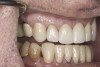
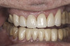
Based on examination of the mounted models, it was decided to treat the patient in centric relation and an additive equilibration approach was used to idealize the planes of occlusion. On the articulator, the patient’s first point of contact in centric relation was the starting point and an additive equilibration technique was implemented, building the teeth to meet ideally at the patient’s first point of contact. A diagnostic wax-up was completed on the mounted models to determine where the anterior and posterior teeth needed to be in space. A template from the wax-up was used to fabricate the anterior provisional restorations. The anterior teeth were restored with full-coverage restorations, creating ideal stops between the lower incisal edges and upper cingulums. The maxillary incisal edges were placed to provide proper phonetics and an acceptable esthetic outcome, while still working within the envelope of function. All of these parameters are worked out in the provisionals prior to moving forward with definitive restorations. With the anterior provisionals in place, posterior composite tops were bonded to his existing dentition, creating ideal centric stops on the posterior teeth. The anterior and canine guidance was developed, resulting in immediate separation of the posterior teeth during all excursive movements (Figure 17 through Figure 28). Once the author and patient were happy with the function, esthetics, phonetics, and comfort of the provisionals, an impression of the approved provisionals was taken and sent to the laboratory to use as a guide in the fabrication of the final anterior crowns. With the final restorations delivered, the patient was extremely happy with his new smile. Most importantly, proper anterior and canine guidance in harmony with his envelope of function was achieved, which resulted in comfortable function and immediate separation of the posterior teeth during protrusive and lateral excursions (Figure 29 through Figure 42).
Conclusion
The author realizes that this case is not considered ideal. Significant compromises were made to the treatment plan because of the wishes of the patient, and the esthetics could have been greatly improved. The final result was an improved smile and a properly functioning masticatory system. This system was ideally planned preoperatively using the proven principles of proper occlusion that have been developed and tested over the years. The final design was then transferred to the patient’s mouth so he could experience comfortable, long-lasting function while enjoying his new smile.
Disclosure
The author has no relevant financial relationships to disclose.
References
1. Turp JC, Greene CS, Strub JR. Dental occlusion: a critical reflection on past, present and future concepts. J Oral Rehabil. 2008;35(6):446-453.
2. Christensen GJ. Abnormal occlusion conditions: a forgotten part of dentistry. J Amer Dent Assoc. 1995;126(12)1667-1668.
3. Parker MW. The significance of occlusion in restorative dentistry. Dent Clin North Am. 1993;37(3):341-351.
4. Wilkins A. The Way of the Superior Dentist: Connecting with Patients, Creating Abundance, and Cultivating Your Passion. North Charleston, SC: CreateSpace Independent Publishing Platform; 2014.
5. Gibbs CH, Messerman T, Reswick JB, Derda HJ. Functional movements of the mandible. J Prosthet Dent. 1971;26(6):604-620.
6. Dawson PE. Functional Occlusion: From TMJ to Smile Design. St. Louis, MO: Mosby; 2006.
7. Tarantola GJ, Becker IM, Gremillion H. The reproducibility of centric relation: a clinical approach. J Am Dent Assoc. 1997;128(9):1245-1251.
8. McKee JR. Comparing condylar positions achieved through bimanual manipulation to condylar positions achieved through masticatory muscle contraction against an anterior deprogrammer: a pilot study. J Prosthet Dent. 2005;94(4):389-393.
9. Owens SE Jr, Lehr RP Jr, Biggs NL. The functional significance of centric relation as demonstrated by electromyography of the lateral pterygoid muscles. J Prosthet Dent. 1975;33(1):5-9.
10. Jung SA, Han KS. Effect of slide in centric relation and head posture on occlusal contact and masticatory muscle activity. Oral Surg Oral Med Oral Pathol Oral Radiol Endod. 2005;99(4):443.
11. Geering AH. Occlusal interferences and functional disturbances of the masticatory system. J Clin Periodontol. 1974;1(2):112-119.
12. Murray GM, Orfanos T, Chan JY, et al. Electromyographic activity of the human lateral pterygoid muscle during contralateral and protrusive jaw movements. Arch Oral Biol. 1999;44(3):269-285.
13. Bhutada MK, Phanachet I, Whittle T, et al. Activity of superior head of human lateral pterygoid increases with increases in contralateral and protrusive jaw displacement. Eur J Oral Sci. 2007;115(4):257-264.
14. Matsunaga K, Usui A, Yamaguchi K, Akita K. An anatomical study of the muscles that attach to the articular disc of the temporomandibular joint. Clin Anat. 2009;22(8):932-940.
15. Aoki S, Uchida S, Inoue H. Fatigue-related changes in discharge patterns of motor units in the inferior head of the lateral pterygoid muscle in humans. Arch Oral Biol. 2005;50(8):727-737.
16. Lopes SL, Costa AL, Gamba Tde O, et al. Lateral pterygoid muscle volume and migraine in patients with temporomandibular disorders. Imaging Sci Dent. 2015;45(1):1-5.
17. Iwasaki LR, Liu H, Gonzalez YM, et al. Modeling of muscle forces in humans with and without temporomandibular joint disorders. Ortho Craniofac Res. 2015;18(Suppl 1):170-179.
18. Franks AS. Masticatory muscle hyperactivity and temporomandibular joint dysfunction. J Prosthet Dent. 1965;15(6):1122-1131.
19. Dawson PE. Evaluation, Diagnosis, and Treatment of Occlusal Problems. St. Louis, MO: Mosby; 1989.
20. Ramfjord S, Ash MM. Occlusion. Philadelphia, PA: W.B. Saunders Company; 1983.
21. Eberhard L, Braun S, Wirth A, et al. The effect of experimental balancing interferences on masticatory performance. J Oral Rehabil. 2014;41(5):346-352.
22. Williamson EH, Lundquist DO. Anterior guidance: its effect on electromyographic activity of the temporal and masseter muscles. J Prosthet Dent. 1983;49(6):816-823.
23. Kerstein RB, Radke J. Masseter and temporalis excursive hyperactivity decreased by measured anterior guidance development. Cranio. 2012;30(4):243-254.
24. Lundeen HC, Gibbs CH. The Function of Teeth: The Physiology of Mandibular Function Related to Occlusal Form and Esthetics. Gainesville, FL: L and G Publishers; 2005.
25. Hess L. Management of the incisal edge. Inside Dentistry. 2013;9(10)58-64.
26. Cranham J. The horizontal position of the maxillary incisal edge: the key to optimum esthetics, phonetics, and function. Contemporary Esthetics and Restorative Practice. 2006:10(2):22-24.
About the Author
Matthew D. Wolfe, DDS
Associate Faculty
The Dawson Academy
St. Petersburg, Florida
Private Practice
Rochester Hills, Michigan
![(1.) Lateral view of cross-section through the TMJ: [1] Posterior slope of the eminentia; [2] condyle; [3] disc; [4] superior lateral pterygoid muscle; [5] inferior lateral pterygoid muscle; [6] synovial tissue; [7] retrodiscal
tissue; [8] posterior ligamentous attachment of disc to condyle.](/media/thumbnail/27829)